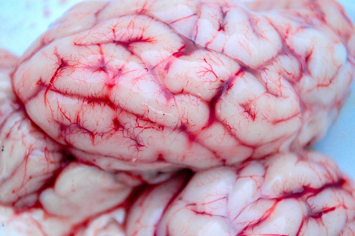

A 3D model of the human brain helps with gaining new insights into neurological abnormalities. In doing so, the model accurately mimics the structure and function of the human brain, and should help in early detection of neurological abnormalities.
The 3D model was developed by researchers at South Korean university POSTECH. Recovery is little to no in neurodegenerative diseases such as Alzheimer's and Parkinson's. This makes early detection and prediction of such diseases of great importance. In practice, however, this is complex, partly due to the complexity of the human brain.
With their model, researchers hope to provide relief. The model should help understand the human brain's response to a variety of conditions in the laboratory. For this purpose, researchers have long been working with two-dimensional cell structures and stem cell-based organoids. In practice, however, both offer limited possibilities for reproducing the complex architecture and function of the human brain.
Bioengineered Neural Network
The new 3D model is a so-called Bioengineered Neural Network (BENN). This is an artificial model of the human brain built layer by layer. The model uses biomimetic compartmentalisation. One part of the model consists of so-called grey matter, which comprises neuronal cell bodies. A second part comprises white matter, which consists of aligned axons that act as information highways that facilitate signal transmission.
The researchers applied electrical stimulation to guide the axonal growth of neurons in a specific direction. Thus, they promoted the formation of aligned and interconnected neural pathways. This led to a functional neural network very similar to the brain's original signal transmission architecture. Real-time monitoring of calcium ion flux confirms that the BENN model exhibits electro-physiological responses analogous to those observed in actual brain tissue, the researchers report
Effects of alcohol exposure investigated
The team also used the BENN platform to investigate the effects of alcohol exposure on brain function. In doing so, the model was treated daily for three weeks with ethanol at a concentration of 0.03%, which the researchers say is representative of moderate social drinking. In the grey matter area, they observed increased levels of Alzheimer's-related proteins, including amyloid-beta and tau. In the white matter, they identified significant morphological changes in neural fibres, including swelling and deformation.
The propagation of neural signals also showed remarkable attenuation. This study is the first to visualise and quantify region-specific neurotoxic responses to alcohol in real-time using a biotechnological brain model.
'Important step forward'
"This model enables high-quality analysis of neural connectivity and electro-physiological responses, which were previously difficult to observe. It has considerable potential for early detection of diseases and accurate prediction of therapeutic outcomes at the preclinical stage," said POSTECH professor Dong-Woo Cho. Professor Jinah Jang added: "This research marks an important step forward in our ability to investigate the early pathological events of brain diseases in a laboratory setting."
The study results were published by researchers in the International Journal of Extreme Manufacturing and can be found here.
Image: aziz enn via Pixabay